Fibroid embolization before pregnancy, with ovarian protection
In a woman in her 30s, rapidly growing fibroids were causing pressure and bleeding symptoms. Fibroid embolization was performed to preserve the uterus in a patient wishing to conceive in the future. The left uterine–ovarian artery collateral was closed with a superselective coil, preventing particle passage into the ovarian circulation; at five months, the symptoms had completely regressed, enhancement had disappeared on MRI, and the fibroids had shrunk by approximately 60–70%.
Rapidly growing fibroids, pressure and bleeding symptoms
A woman in her 30s presented with a feeling of pressure and bleeding symptoms caused by rapidly growing fibroids. She wished to preserve her uterus and was planning a pregnancy in the future.
For this reason, the treatment decision was not based on the size of the fibroids alone. The severity of the symptoms, growth tendency, imaging findings and the patient’s reproductive plans were assessed together.
Multiple, markedly enhancing fibroids on imaging
Pelvic MRI showed multiple fibroids enlarging the uterus. The marked enhancement of the fibroids indicated active arterial blood supply.
Selective angiography during the procedure showed that the fibroids were supplied mainly by the left uterine artery, while the contribution from the right uterine artery was more limited.
A minimally invasive treatment option that preserves the uterus
The decision to perform uterine artery embolization was made after considering together the patient’s wish to preserve her uterus, the severity of her symptoms and the imaging features of the fibroids.
In patients wishing to conceive, the decision to perform fibroid embolization should be made on an individual basis. The uncertainties of possible surgical and interventional options regarding fertility should be discussed with the patient; the procedure should not be presented as a guarantee of pregnancy.
Hypogastric plexus block and bilateral embolization
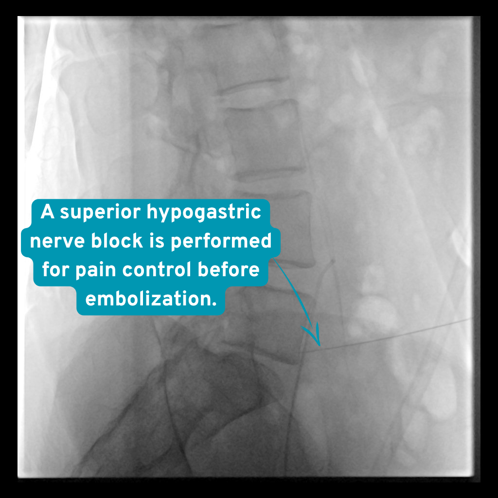
A hypogastric nerve/plexus block was performed first to help reduce post-procedural pain. The left uterine artery was then catheterised superselectively. The dominant fibroid supply was confirmed to be from the left side, and embolization with microsphere particles was started.
On an interim control run, a collateral vessel became visible between the left uterine artery and the left ovarian artery. This vessel was catheterised superselectively with a microcatheter and closed with a coil. After confirming that the route for non-target particle passage into the ovarian circulation had been closed, embolization of the left uterine artery was completed.
The right uterine artery was then catheterised, and the remaining fibroid blood supply was embolized with microspheres. Once the intended loss of blood supply was seen on bilateral control angiograms, the procedure was ended.
The story on DSA: seven steps from dominant left-sided supply to ovarian collateral protection
-
What you see Marked and dense enhancement arising from the left uterine artery is seen in the fibroid bed.
Why it matters This appearance showed that the dominant feeder to the fibroids was on the left, supporting starting embolization from this side.
During the procedure The initial DSA shows that the fibroids are supplied predominantly by the left uterine artery. Initial DSA series obtained after selective catheterisation of the left uterine artery. -
What you see A collateral vascular connection becomes visible between the circulation of the left uterine artery and the left ovarian artery.
Why it matters The possibility that microspheres could pass into the ovarian circulation through this connection required an additional precaution against non-target embolization in a patient wishing to conceive.
During the procedure The collateral connection between the left uterine artery and the left ovarian artery becomes visible during the interim check. Interim control DSA series performed while embolization of the left uterine artery was ongoing. -
What you see A small coil is seen being placed in a controlled manner through a microcatheter advanced into the collateral vessel.
Why it matters The coil was used to close the collateral route in order to reduce the risk of non-target passage of particles into the ovarian circulation while embolization continued.
During the procedure The uterine–ovarian collateral is catheterised superselectively and closed with a coil. The stage at which the uterine–ovarian collateral is catheterised superselectively with a microcatheter and closed with a coil. -
What you see The previously opacified ovarian collateral connection is no longer visible while the coil remains in place.
Why it matters After confirming that the route for non-target passage had been closed, embolization of the left uterine artery was continued in a more controlled manner.
During the procedure The disappearance of the ovarian connection is confirmed after coil placement, and embolization on the left side continues. Control DSA series obtained from the left uterine artery after collateral coil embolization. -
What you see The dense fibroid enhancement seen initially is no longer visible, while the coil material can be seen.
Why it matters This appearance showed that the intended embolization endpoint had been reached on the left side.
After the procedure The blood supply in the fibroid bed is seen to have stopped at the end of left uterine artery embolization. Control angiogram obtained after completion of left uterine artery embolization. -
What you see More limited vascular supply extending from the right uterine artery to the fibroid bed is seen compared with the left side.
Why it matters Although the dominant feeder to the fibroids was on the left, treating the remaining right-sided blood supply completed the bilateral embolization.
During the procedure More limited fibroid blood supply is seen on right uterine artery DSA; the dominant feeder was on the left. DSA series in which the right uterine artery was assessed selectively after completion of the left side. -
What you see Flow in the right uterine artery branches is slowing, and no marked residual enhancement is seen in the fibroid bed.
Why it matters This final series confirmed that the right side had also been embolized to the intended level, and the procedure was ended.
After the procedure No marked residual blood supply is seen in the fibroid bed after right uterine artery embolization. Final control series obtained after completion of right uterine artery embolization.
Balancing safety when pregnancy is desired
Closing the vessel with a coil does not guarantee preservation of fertility; the aim here was to reduce an identified risk of unintended particle passage into the ovarian circulation through the observed collateral. Pregnancy is possible after fibroid embolization, but in every patient planning a pregnancy, ovarian reserve, the location of the fibroids and alternative treatments should be assessed on a multidisciplinary basis.
Comparison of MRI before the procedure and at five months
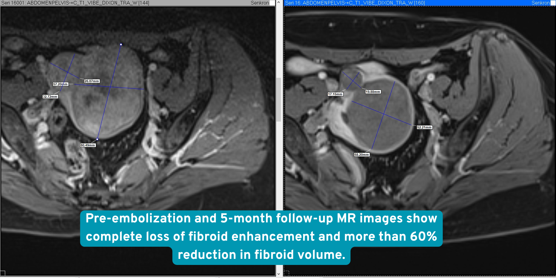
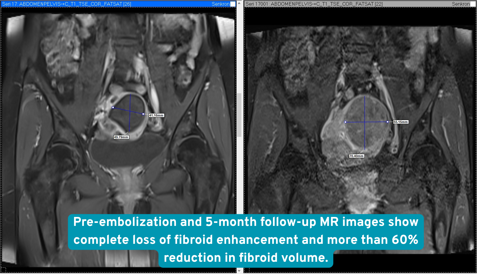
One-day stay, marked response at five months
The patient was kept under observation for one day after the procedure and was then discharged. At the five-month follow-up, the pressure and bleeding symptoms were seen to have completely regressed.
No residual enhancement was detected in the fibroids on follow-up MRI; this finding was consistent with complete devascularisation. A reduction of approximately 60–70% in fibroid volume was observed.
What this case teaches
In fibroid embolization, interim checks performed as embolization progresses are important, not only the initial angiogram. As the flow balance changes, uterine–ovarian collaterals that were not visible initially may become prominent.
Recognising this connection in time and closing it when necessary with superselective coil embolization may reduce the risk of non-target embolization and allow uterine artery embolization to be completed in a controlled manner.
What this means for the patient
Uterine artery embolization is a minimally invasive option that can reduce fibroid-related bleeding and pressure symptoms in suitable patients without removing the uterus. When both uterine arteries and possible ovarian connections are assessed in detail during treatment, the procedure can be adapted to the individual vascular anatomy.
The decision is not standardised in patients planning a pregnancy. Expectations, alternatives and uncertainties regarding fertility should be considered together with specialists in interventional radiology and gynaecology.
Risks and alternatives
Possible risks
Cramping and pain are common after treatment. Bleeding, infection, artery or uterine injury, ovarian injury or early menopause can occur. The effect on a future pregnancy is uncertain; a rare cancer mistaken for a fibroid could be diagnosed late.
Other options
Medication, myomectomy or hysterectomy, depending on symptoms, fibroid anatomy and pregnancy plans. Discuss fertility goals with a gynaecologist before deciding.
Clinical source: ACOG
This is one patient. Imaging is anonymised and published with that patient’s written consent. Individual results vary, and nothing here is a prediction of your own outcome or a substitute for a consultation.