Uterine fibroid embolization to preserve the uterus
A woman in her 30s had symptoms of pressure, frequent urination, pain and bleeding due to fibroids. Uterine artery embolization was performed in line with her wish to preserve her uterus; at the six-month MRI, complete devascularization and more than 75% volume reduction were observed.
Symptoms of pressure, frequent urination, pain and bleeding
A woman in her 30s presented with a marked sensation of pressure, frequent urination, pain and bleeding due to fibroids. The symptoms were significantly affecting her daily life.
The patient did not want her uterus to be removed and did not prefer surgical treatment. She was therefore assessed for uterus-preserving minimally invasive options.
Large and markedly vascularised fibroid on MRI
Pelvic MRI showed a large fibroid causing marked enlargement of the uterus and pressure on surrounding structures. On contrast-enhanced sequences, the fibroid was seen to be viable and markedly vascularised.
When the clinical symptoms and imaging findings were assessed together, the fibroid was considered to be consistent with the symptoms of pressure, frequent urination, pain and bleeding.
A non-surgical uterus-preserving treatment option
The severity of the symptoms, the size and vascularity of the fibroid, and the patient’s wish to preserve her uterus and avoid surgery were assessed together. After the suitable options had been discussed, uterine artery embolization was chosen.
The aim of treatment was to control bleeding by reducing arterial flow to the fibroid and to reduce pressure symptoms as the fibroid became smaller over time.
Bilateral uterine artery embolization
The procedure was performed by selectively catheterising both uterine arteries under angiographic guidance. The initial DSA series confirmed marked arterial supply extending to the fibroid.
The uterine arteries were embolized in a controlled manner with microsphere particles. The procedure was concluded when no marked residual blood supply to the fibroid was seen on the final control series on the right and left sides. Pain control and clinical follow-up were planned after the procedure.
The procedure on DSA: three steps from vascularisation to devascularization
-
What you see Marked contrast enhancement originating from the left uterine artery is seen in the fibroid bed.
Why it matters This appearance confirmed active arterial supply and showed the target for embolization.
During the procedure Marked vascularisation extending from the left uterine artery to the fibroid is seen on the initial DSA. Selective DSA series obtained from the left uterine artery before embolization. -
What you see The marked fibroid enhancement seen initially is observed to have disappeared.
Why it matters This appearance showed that the intended embolization endpoint on the left had been reached.
After the procedure No marked vascularisation is seen in the fibroid bed after embolization of the left uterine artery. Control DSA series obtained after completion of left uterine artery embolization. -
What you see No marked residual contrast enhancement of the fibroid is seen on injection of the right uterine artery.
Why it matters Completion of bilateral embolization was confirmed and the procedure was concluded.
After the procedure No marked residual vascularisation of the fibroid is seen after embolization of the right uterine artery. Control DSA series obtained after completion of right uterine artery embolization.
Comparison before the procedure and at six months on MRI
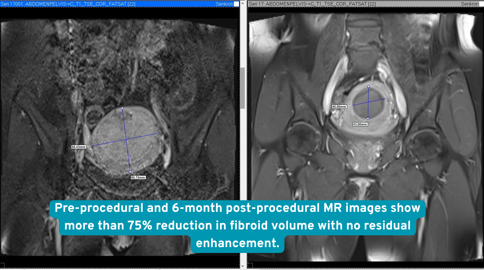
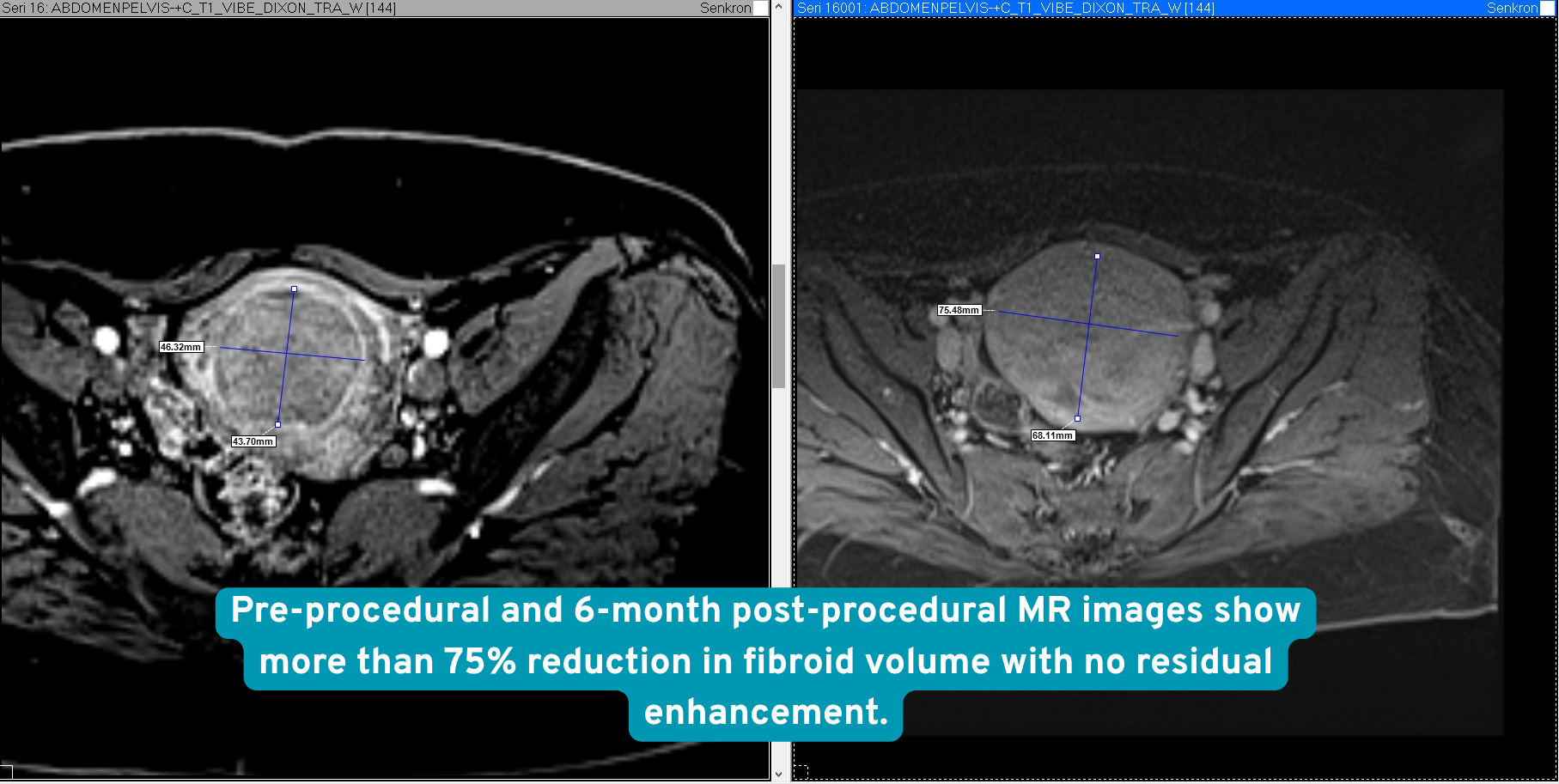
One-day admission, with marked clinical and radiological response at six months
The patient was kept under observation for one day after the procedure and was then discharged. At the six-month follow-up, the symptoms of pressure, frequent urination, pain and bleeding were seen to have regressed markedly.
The follow-up MRI showed that the fibroid was completely devascularized and that its volume had decreased by more than 75%.
What this case teaches
In large fibroids, pressure effects may cause not only pain but also bladder symptoms and frequent urination. Treatment success should therefore be assessed not only by the reduction in bleeding, but also together with changes in pressure symptoms and follow-up MRI findings.
In this case, after bilateral uterine artery embolization, both marked improvement in the clinical symptoms and complete devascularization with more than 75% reduction in volume on the six-month MRI were observed.
What this means for the patient
Uterine artery embolization is a minimally invasive treatment option that, in suitable patients, can reduce bleeding and pressure symptoms caused by fibroids without removing the uterus or performing open surgery.
For each patient, the decision should be made after considering together the number, size and location of the fibroids, the symptoms, age, coexisting conditions and personal expectations.
Risks and alternatives
Possible risks
Cramping and pain are common after treatment. Bleeding, infection, artery or uterine injury, ovarian injury or early menopause can occur. The effect on a future pregnancy is uncertain; a rare cancer mistaken for a fibroid could be diagnosed late.
Other options
Medication, myomectomy or hysterectomy, depending on symptoms, fibroid anatomy and pregnancy plans. Discuss fertility goals with a gynaecologist before deciding.
Clinical source: ACOG
This is one patient. Imaging is anonymised and published with that patient’s written consent. Individual results vary, and nothing here is a prediction of your own outcome or a substitute for a consultation.