Bilateral varicocele embolization through one femoral access
A patient in their 30s had bilateral varicocele accompanied by pain, a low sperm count and reduced sperm motility. The left side was embolized first, followed by the right, through a single femoral access. At Doppler follow-up at one, three and six months, the vein diameters were normal and no reflux was seen; the pain had resolved, and a semen analysis was requested for follow-up.
Bilateral Varicocele with Pain and Impaired Semen Parameters
A male patient in his 30s had varicocele on both the left and right sides. His complaint of scrotal pain was accompanied by a reduced sperm count and reduced motility on his previous semen analysis.
For this reason, the assessment did not focus only on the source of the pain. The anatomy of the venous reflux on both sides, the existing semen findings and whether both sides could be treated in the same session were considered together.
Doppler and Angiography: Reflux on Both Sides
Pre-procedure assessment showed bilateral varicocele. Colour Doppler was used to assess dilated testicular veins and venous reflux, while also allowing comparison with later follow-up examinations.
Venography performed during the procedure showed marked reflux towards the varicocele, first in the left gonadal vein and then on the right. The reflux pathway on each side was therefore mapped separately during the same procedure.
Why Was Treatment of Both Sides Planned in the Same Session?
The patient’s varicocele was bilateral; in addition to pain, the semen analysis showed a reduced sperm count and reduced motility. These findings were considered together, and embolization of the left and right sides in the same session was planned if suitable venous anatomy could be reached.
The aim was not simply to occlude the two main gonadal veins. The main and collateral pathways carrying blood towards the varicocele were to be identified on each side, open collateral vessels were to be sought, and the procedure was to be completed only when reflux had ceased on control imaging.
Left First, Then Right, Through a Single Femoral Access
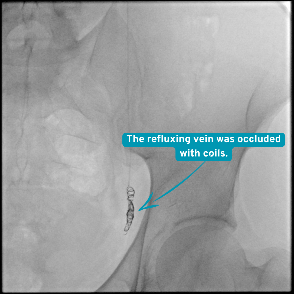
The procedure began with a single needle access through the femoral vein. The catheter was advanced first into the left gonadal vein. Initial venography showed marked reflux towards the varicocele, and the main reflux pathway was occluded with coils.
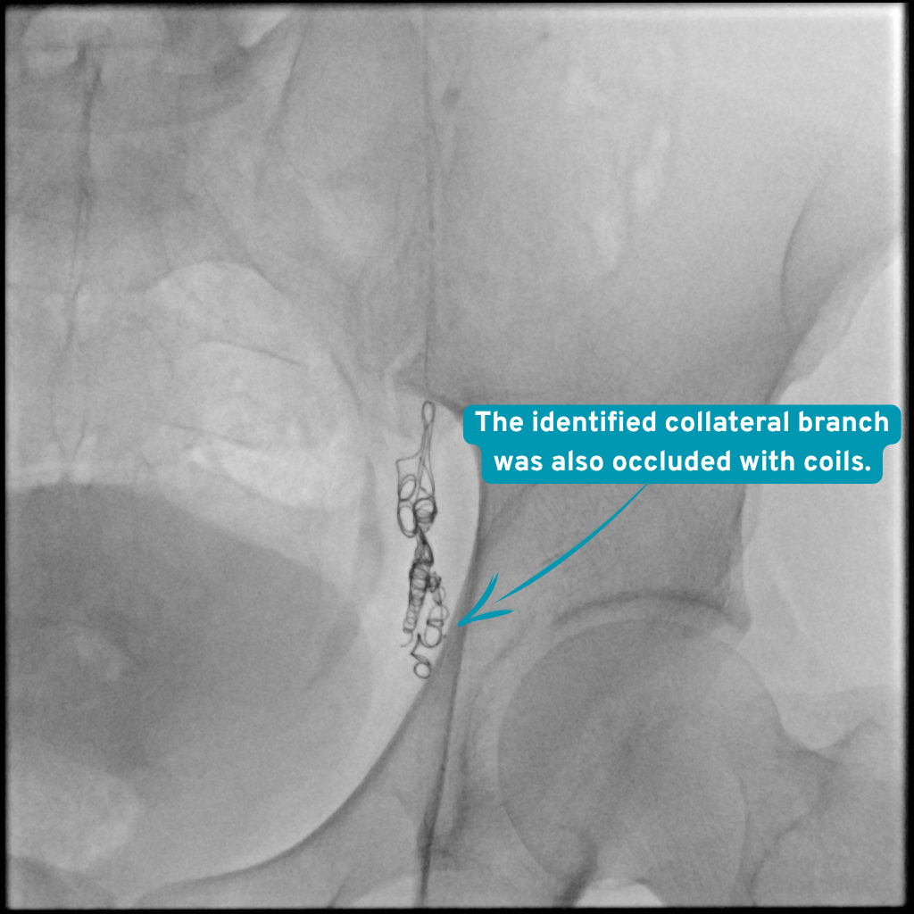
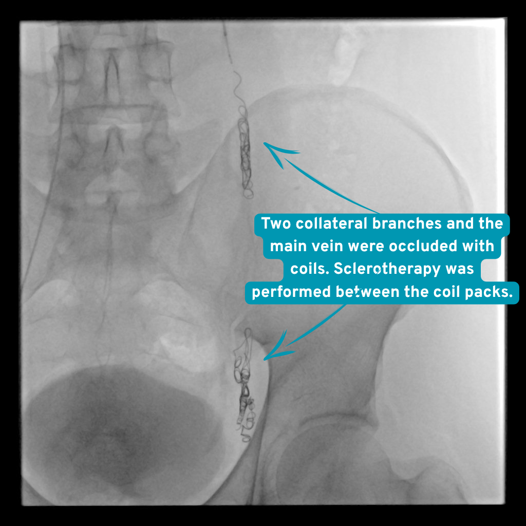
The first control image and the Valsalva manoeuvre appeared to show that leakage through the main pathway had stopped. However, an additional series obtained from different angles and with different manoeuvres showed that a collateral branch had opened and was maintaining the reflux. This collateral pathway was also occluded with coils; sclerotherapy was applied to the target segments between the coil packs. On the final control from different angles on the left, cessation of reflux was confirmed.
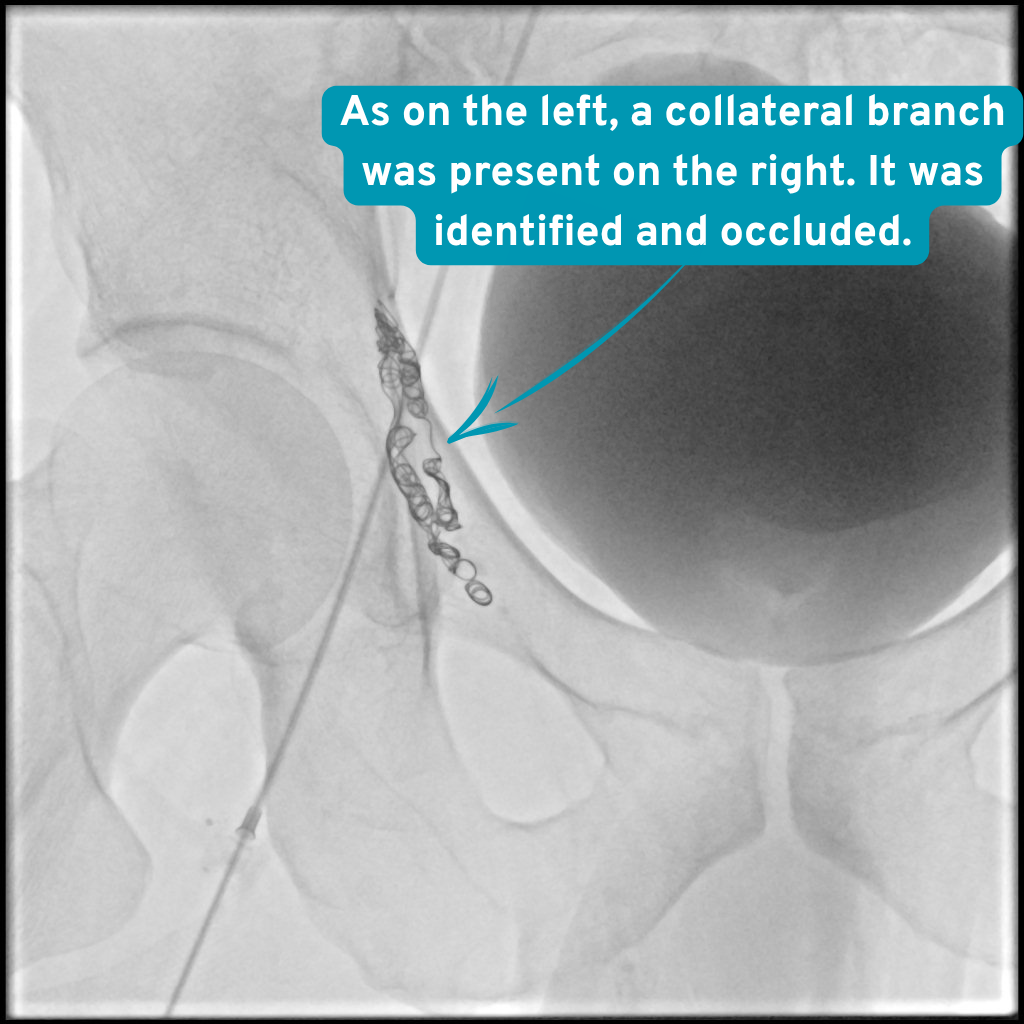
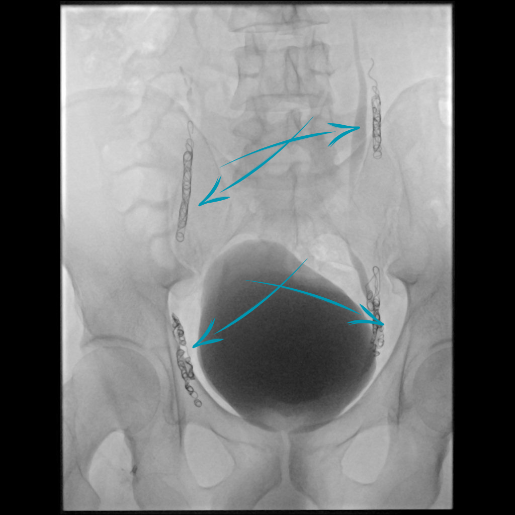
The right gonadal venous system was then entered while maintaining the same femoral access. Marked reflux was also demonstrated on the right. The leaking venous pathways were occluded with coils and sclerotherapy; on the final control images, the right-sided reflux was also seen to have ended.
The Venographic Story: Seven Steps from Left to Right
-
What you see Contrast is seen flowing backwards from the left gonadal vein downwards, towards the varicocele.
Why it matters The initial image shows the location of the reflux pathway on the left and which venous route the treatment should target.
During the procedure Marked reflux towards the varicocele is seen in the left gonadal vein. The first venography series obtained after reaching the left gonadal vein through the femoral access. -
What you see The coil pack in the leaking vessel and the position of the catheter within the vein are seen.
Why it matters The coils provide mechanical occlusion in the main reflux pathway; the area is then checked separately for any remaining open collateral pathway.
During the procedure The main pathway carrying reflux on the left was occluded with coils. The first embolization step for the main venous pathway carrying the reflux. -
What you see In this projection, no reflux towards the distal part is seen from the main pathway occluded with coils.
Why it matters A single control image may not prove that all collaterals have been occluded; assessment from different angles was therefore continued.
During the procedure Reflux in the main pathway had stopped; additional checks were performed before ending the procedure. The control series obtained with the Valsalva manoeuvre after the first coil application. -
What you see Although the main pathway is closed, a collateral branch is seen to fill and continue the reflux.
Why it matters This image showed that the first technical step was not sufficient and that the open collateral pathway also needed to be targeted.
During the procedure A different angle made the collateral vessel maintaining reflux on the left visible. Additional venography obtained after changing the projection and manoeuvre following the initial control. -
What you see During contrast injection, no further venous filling towards the varicocele is seen on the left.
Why it matters The angiographic endpoint on the left was reached after both the main and collateral pathways had been checked.
During the procedure Cessation of reflux was confirmed on final left venography. The final left series obtained from different angles and with different manoeuvres after the collateral pathway had also been occluded. -
What you see Contrast is seen to flow markedly backwards towards the varicocele on the right as well.
Why it matters In bilateral treatment, the right side also needs to be assessed separately, with its own venous anatomy.
During the procedure Marked reflux was demonstrated on right gonadal venography. After the left side was completed, the right gonadal venous system was entered through the same femoral access. -
What you see The coil packs are seen, with no continuing reflux towards the varicocele.
Why it matters This control shows that the right side was also completed and that a separate endpoint was reached for each side during the same session.
After the procedure Reflux on the right ended after coils and sclerotherapy. The final control series obtained after treatment of the leaking venous pathways on the right.
Main and Collateral Pathways Occluded with Coils
Doppler Follow-up at One, Three and Six Months
The patient attended follow-up at one, three and six months. At all three visits, colour Doppler ultrasonography showed that the testicular vein diameters had returned to within normal limits and that no reflux was seen. The patient’s pain had resolved.
As the sperm count and motility had initially been reduced, a follow-up semen analysis was requested. At the time this draft was prepared, the new semen analysis result was not yet available; therefore, no result was reported regarding the extent to which the semen parameters had changed.
What this case teaches
Even when treatment in the same session is planned for bilateral varicocele, the right and left sides are not treated as a single venous system. The gonadal vein, collateral connections and control venography of each side are assessed separately.
The most instructive point in this case was that, although no leakage was seen in the first control of the main left pathway, collateral reflux appeared with a different angle and manoeuvre. The technical endpoint was determined not simply by placing the coils, but by demonstrating that no open collateral pathway remained and that reflux had ceased.
What this means for the patient
Bilateral varicocele does not mean that both sides will necessarily be treated in the same session in every patient. The decision is made by considering the symptoms, examination, Doppler findings, semen assessment and the individual venous anatomy together.
In this patient, resolution of the pain and the absence of reflux together with normal vein diameters at Doppler follow-up at one, three and six months were favourable follow-up findings. However, changes in sperm count and motility cannot be inferred from Doppler; the follow-up semen analysis requested for this purpose must be assessed separately. These results relate to a single patient and do not constitute a guarantee of outcomes for other patients.
Risks and alternatives
Possible risks
Temporary discomfort, bruising, minor bleeding or infection can occur. Rarely, an embolic coil or liquid may move; the varicocele can persist or recur and further treatment may be needed.
Other options
Observation when symptoms are mild, or surgical repair after a fertility and urology assessment.
Clinical source: CIRSE
This is one patient. Imaging is anonymised and published with that patient’s written consent. Individual results vary, and nothing here is a prediction of your own outcome or a substitute for a consultation.