Nutcracker syndrome: ovarian vein embolization and renal stent
In a woman in her 30s, flank and abdominal pain were accompanied by a dilated left ovarian vein, pelvic varices, mild left kidney oedema, compression of the left renal vein between the aorta and superior mesenteric artery, and proteinuria. The left ovarian vein was embolized first in the same session, followed by stent placement in the left renal vein; the pain decreased markedly within a few weeks and the assessment of kidney function returned to normal.
Presentation with flank pain, abdominal pain and proteinuria
A woman in her 30s presented with flank pain and abdominal pain. During assessment of her symptoms, not only the pelvic vessels but also the venous drainage of the left kidney and findings related to kidney function were examined together.
Proteinuria identified in laboratory testing was one of the clinical findings suggesting that the compression of the left renal vein seen on imaging might not be only an anatomical appearance.
Left renal vein compression, dilated ovarian vein and pelvic varices
Imaging showed that the left ovarian vein was markedly dilated and that widespread varicose veins were present in the pelvis. The left kidney appeared mildly oedematous. The left renal vein was seen to be compressed between the aorta and the superior mesenteric artery, with marked stenosis in this segment.
Venography showed the stenosis at the origin of the left renal vein together with the dilated left ovarian vein. On selective imaging, reflux of contrast along the left ovarian vein was seen filling the pelvic variceal bed around the uterus.
Why was a two-stage treatment planned?
There were two interconnected venous problems in this case. Reflux into the pelvis through the dilated left ovarian vein was filling the pelvic variceal bed associated with the pain. Above this, stenosis of the left renal vein in the aortomesenteric segment was the main point of compression affecting renal venous drainage.
Flank and abdominal pain, proteinuria, mild left kidney oedema and the reflux shown on venography were assessed together, and a two-stage plan to be carried out in a single session was made. The first stage aimed to embolize the left ovarian vein reflux and pelvic varices, while the second stage aimed to open the left renal vein stenosis with a venous stent.
First-stage embolization, second-stage renal vein stenting
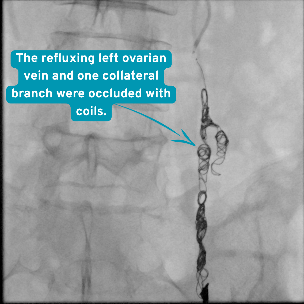
The first stage targeted the leaking left ovarian vein and the pelvic variceal bed. The left ovarian vein and a collateral branch involved in the reflux were closed with coils; polidocanol (Aethoxysklerol) was applied to the target venous segments as a sclerosant. Control venography showed that reflux into the pelvic varices had stopped.
After embolization was completed, the left renal vein was reassessed. Although leakage towards the ovarian vein was no longer seen, the stenosis between the aorta and the superior mesenteric artery persisted. In the second stage of the same session, a venous stent was placed in the narrowed left renal vein.
The story on venography: seven steps from stenosis to embolization and stenting
-
What you see The left renal vein is seen to be markedly narrowed in the aortomesenteric segment, while the left ovarian vein fills as a dilated outflow pathway.
Why it matters Seeing both the renal vein compression point and the dilated ovarian vein carrying pressure towards the pelvis in the same image established the anatomical basis for the two-target plan.
During the procedure The initial venogram shows left renal vein stenosis together with a markedly dilated left ovarian vein. Initial venography showing the left renal venous anatomy and collateral pathway before treatment. -
What you see Contrast is seen flowing back down along the left ovarian vein and filling the pelvic variceal bed around the uterus.
Why it matters This series directly showed the reflux pathway supplying the pelvic varices and identified the target of the first treatment stage.
During the procedure Reflux from the left ovarian vein is filling the pelvic variceal bed around the uterus. A venography series selectively assessing the dilated left ovarian vein. -
What you see The coils are seen being placed in a controlled manner along the dilated left ovarian vein.
Why it matters The coils provided mechanical closure, while polidocanol was used for a sclerosant effect in the target venous segments.
During the procedure The left ovarian vein is being embolized with a combination of coils and polidocanol. The embolization stage in which the leaking left ovarian vein and associated venous pathways were treated. -
What you see The coil packs are seen, with no continuing reflux filling the pelvic variceal bed around the uterus.
Why it matters This check showed that the angiographic endpoint for the first stage of treatment had been reached.
After the procedure After embolization, reflux into the pelvic varices through the left ovarian vein was completely stopped. Control venography obtained after the left ovarian vein and collateral branch had been closed. -
What you see Reflux towards the left ovarian vein is no longer seen, whereas the stenosis in the aortomesenteric segment of the left renal vein is seen to persist.
Why it matters The first stage removed the reflux; the persistent renal vein stenosis was the target of the stent decision in the second stage.
During the procedure Control venography showing persistent left renal vein stenosis after the pelvic leakage had been closed. A control series reassessing the left renal vein after ovarian vein embolization. -
What you see The venous stent is seen positioned to cover the narrowed segment of the left renal vein.
Why it matters The stent was placed to keep open the narrowed venous segment compressed between the aorta and the superior mesenteric artery.
During the procedure A venous stent is being placed in the aortomesenteric stenotic segment of the left renal vein. A DSA series showing treatment of the left renal vein stenosis with a stent in the second stage of the same session. -
What you see The embolization materials placed along the left ovarian vein are seen, with no reflux into the pelvic variceal bed.
Why it matters The final series confirmed that the ovarian vein reflux targeted in the first stage remained closed at the end of the procedure.
After the procedure At the end of the procedure, reflux in the left ovarian vein was confirmed to have stopped completely. The final venography series obtained after completion of the two-stage treatment.
Appearance of the left ovarian vein and collateral branch after embolization
Marked reduction in pain and normalisation of kidney assessment within a few weeks
Within a few weeks after the procedure, the patient's flank pain and abdominal pain had decreased markedly.
At follow-up assessment, findings related to kidney function had returned to within normal limits. Thus, the clinical improvement in pain was accompanied by improvement in the kidney-related assessment.
What this case teaches
Narrowing of the left renal vein between the aorta and the superior mesenteric artery may also be seen incidentally on imaging. In assessing Nutcracker syndrome, the effect of anatomical compression on the patient's symptoms, urinary and kidney findings, and venous collateral circulation is considered together.
In this case, the pelvic varices were not only an independent problem within the pelvis; the dilated left ovarian vein had become a collateral pathway through which pressure from the left renal vein was directed towards the pelvis. Therefore, rather than embolizing only the pelvic reflux or treating only the renal vein stenosis, both targets were addressed sequentially within the same session.
What this means for the patient
Seeing left renal vein compression or a dilated ovarian vein on imaging does not mean that embolization or stenting is required in every patient. The treatment decision is made by assessing the nature of the symptoms, urinary and kidney findings, pelvic varices, venous reflux and the individual's vascular anatomy together.
In this patient, the marked reduction in pain within a few weeks and normalisation of the kidney function assessment after two-stage treatment were favourable follow-up findings. This result belongs to one patient and does not constitute a guarantee of outcome for others.
Risks and alternatives
Possible risks
Pelvic aching may increase temporarily. Bleeding or infection can occur; rarely, embolic material may move to the lungs. Pain may persist or return, especially if another cause of pelvic pain is present.
Other options
Assessment for other causes of pelvic pain, symptom management and gynaecological treatment where appropriate. Embolization is considered only after the venous diagnosis is supported.
Clinical source: CIRSE
This is one patient. Imaging is anonymised and published with that patient’s written consent. Individual results vary, and nothing here is a prediction of your own outcome or a substitute for a consultation.