Recurrent varicocele embolization after two prior surgeries
In a patient in his late 20s, pain and a visible left varicocele persisted despite two operations and one embolization. Three fine collateral pathways identified on angled venography were closed with coils and polidocanol; the pain had resolved at one month, and no reflux was seen on Doppler follow-up at three and six months.
Persistent symptoms despite three treatments
A male patient in his late 20s presented with symptoms related to left scrotal pain and a varicocele that had become visible from the outside. He had previously undergone two operations for a left varicocele, followed by one embolization. Despite this, he had seen no clear benefit from any of the three procedures; his pain and the appearance of dilated veins had persisted.
This history required not only the question “Is there a varicocele again?” to be addressed, but first an explanation of which venous pathway was allowing reflux to continue despite the previous treatments.
Doppler: diameter above 4 mm, marked reflux
On colour Doppler ultrasound, the diameter of the left varicocele veins exceeded 4 mm. Marked reflux flow accompanied the venous dilatation. Considered together with the clinical symptoms, the findings supported haemodynamically significant venous reflux that was continuing despite the previous interventions.
Why repeat embolization?
Because the patient's pain persisted, the varicocele was visible and marked reflux was confirmed on Doppler, it was decided to remap the venous anatomy angiographically. Because of the previous operations and embolization, the main aim this time was not only to visualise the main gonadal vein, but also to identify fine parallel veins, duplications and collateral connections that could sustain the reflux.
Step-by-step venous mapping through jugular access
The procedure began through jugular venous access, and the left gonadal venous system was reached with a catheter. Although coils from the previous embolization were seen on the initial runs, continued marked reflux was confirmed. To reduce vessel overlap, venography was performed from different angles and with more selective injections.
These runs identified fine collateral pathways running alongside the main vessel. On the check performed after the first pathway was closed, reflux was continuing through other collateral branches. The second and third pathways were then embolized in sequence. Coils were used for mechanical occlusion and polidocanol (Aethoxysklerol) as the sclerosing agent to close the collaterals; after each step, a new check run was performed to assess whether any open escape pathway remained.
The story on venography: seven steps from reflux to complete closure
-
What you see While the coil pack from the previous embolization is seen, contrast is seen flowing back distally towards the varicocele.
Why it matters The presence of coils on the image is not sufficient on its own; treatment effectiveness is assessed by whether the reflux has actually been interrupted.
During the procedure Marked reflux continues on the initial run despite the previous coils. The first series obtained after reaching the left gonadal venous system through jugular venous access. -
What you see Contrast can be seen progressing around the previous coil pack and filling an open venous pathway.
Why it matters Vessels that overlap in a single plane may separate on images from different angles; for this reason, the initial image alone was not considered sufficient.
During the procedure The angled image shows more clearly the reflux continuing around the coils. The same region was examined in a different projection. -
What you see A fine collateral pathway extending outside and parallel to the main gonadal vein fills with contrast.
Why it matters If such a fine collateral pathway remains open, reflux may continue even when the main vessel appears closed.
During the procedure The first collateral escape pathway appears on selective venography. The first fine collateral branch became apparent on more selective and angled venography. -
What you see In a different angle, a second collateral vein adjacent to the previous coil pack fills.
Why it matters Finding one escape pathway does not mean that the search is over; other possible connections must also be sought systematically.
During the procedure The second collateral pathway is distinguished in a different projection. The anatomy was not limited to a single collateral. -
What you see Although flow in the first collateral has stopped, contrast is still seen escaping distally through other collateral pathways.
Why it matters This check showed that the first technical step had been successful, but that the entire source of the reflux had not yet been eliminated.
During the procedure The first collateral pathway was closed; an alternative venous escape pathway continued. An immediate check venogram was performed after the first collateral pathway was embolized. -
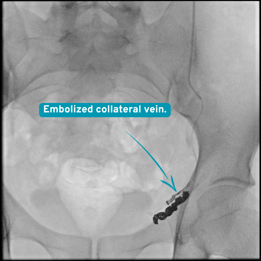
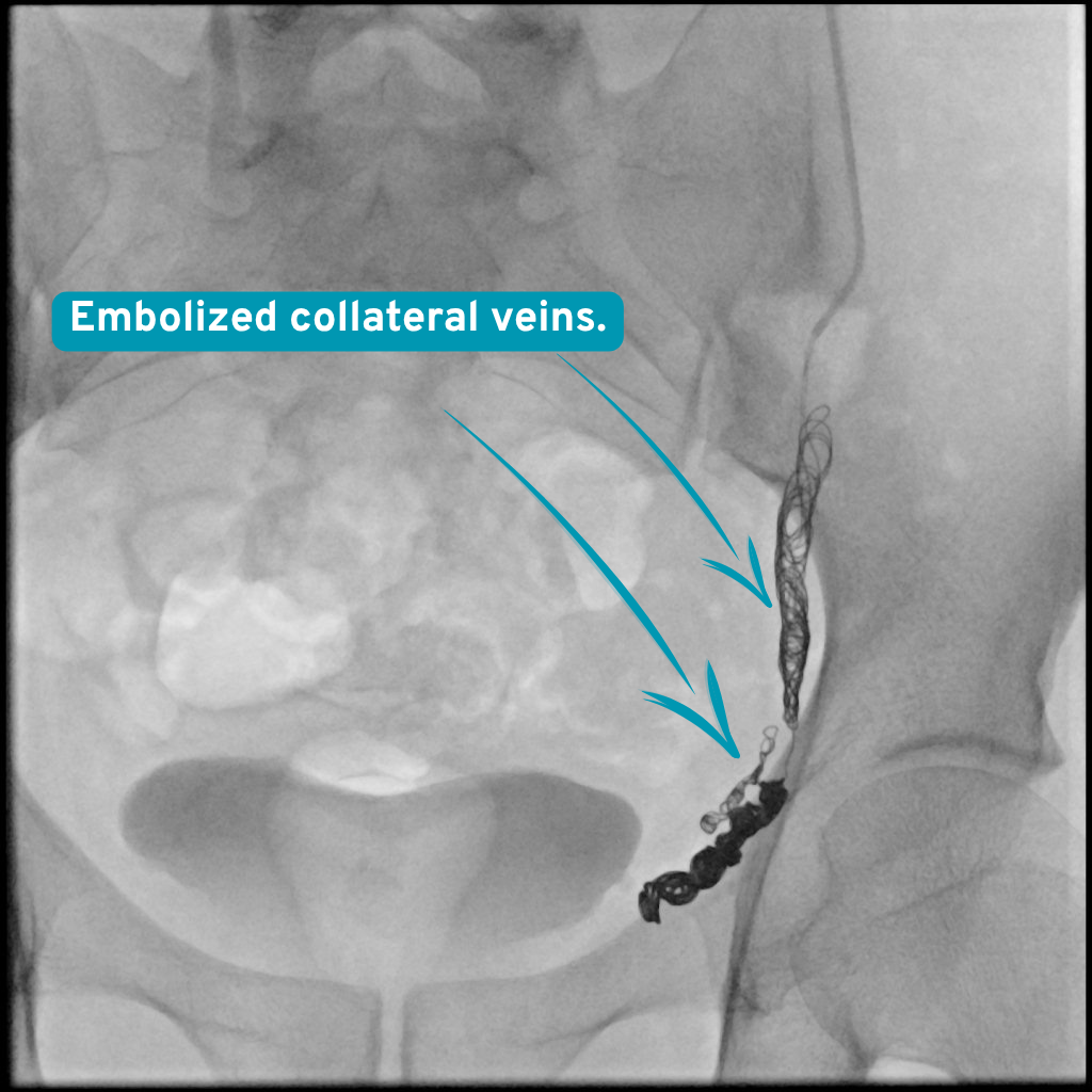
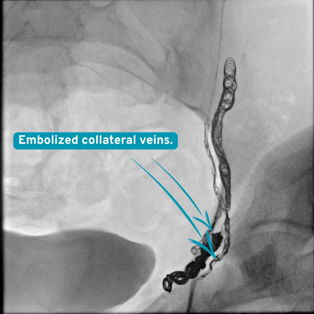
What you see The appearance of the additional collateral pathways identified around the previous coil pack after closure with coils is seen.
Why it matters The aim was not only to disable the visible main vein, but all the connections carrying blood back to the varicocele.
During the procedure The escape network was completed by closing the second and third collateral pathways. Other fine pathways carrying the continuing reflux were also identified and closed using coils and polidocanol (Aethoxysklerol) together. -
What you see During contrast injection, no venous filling towards the distal vessels or reflux towards the varicocele is seen any longer.
Why it matters This image shows that the angiographic endpoint of the procedure had been reached.
After the procedure On the final check, the previous reflux has been completely eliminated. A final venogram was obtained after all the identified collaterals had been closed.
Appearance of the collateral pathways after embolization
Final venography and follow-up
On the final check venogram, reflux towards the varicocele had been completely eliminated. No complication developed during or after the procedure. The patient attended the one-, three- and six-month follow-up appointments regularly.
At one month, his pain had resolved. At the three-month Doppler follow-up, the diameters of the varicocele veins had returned to within normal limits and no reflux was seen. There was also no reflux at the six-month Doppler follow-up; the clinical and imaging improvement was maintained.
What this case teaches
In recurrent varicocele, the cause of failure is not always that the main vessel has reopened. Fine collateral veins or venous duplications may sustain reflux. For this reason, especially in patients who have previously been treated, multi-angle venography, selective catheterisation and a check run after each closure are important.
In this case, the decisive point was to trace the continuing reflux rather than say “the procedure is complete” after the first closure.
What this means for the patient
Having undergone more than one previous treatment does not mean that a new assessment should simply repeat the old procedure. In cases of persistent symptoms, identifying again which vessel is sustaining the problem may guide treatment according to the individual's anatomy. The outcome in this case belongs to one patient; in people with similar symptoms, the decision should be made after considering the clinical examination, Doppler findings and venous anatomy together.
Risks and alternatives
Possible risks
Temporary discomfort, bruising, minor bleeding or infection can occur. Rarely, an embolic coil or liquid may move; the varicocele can persist or recur and further treatment may be needed.
Other options
Observation when symptoms are mild, or surgical repair after a fertility and urology assessment.
Clinical source: CIRSE
This is one patient. Imaging is anonymised and published with that patient’s written consent. Individual results vary, and nothing here is a prediction of your own outcome or a substitute for a consultation.