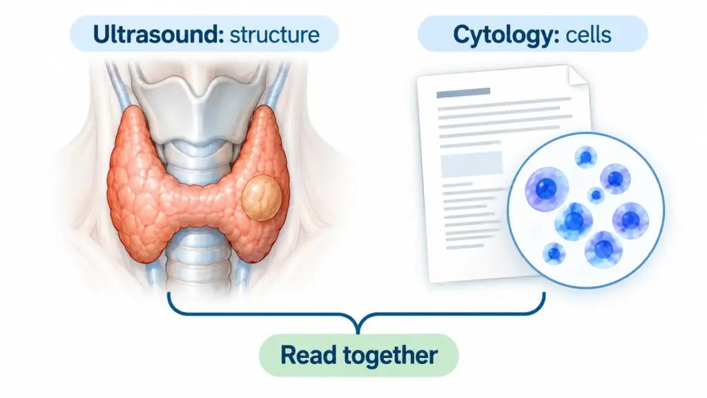
Bethesda categories describe how a pathologist interprets cells collected from a thyroid nodule by fine-needle aspiration, or FNA. Category I is nondiagnostic, II benign, III atypia of undetermined significance, IV follicular neoplasm, V suspicious for malignancy and VI malignant. The category helps guide the next step, but it does not replace the full report and clinical assessment.[1]
A Bethesda number is not a thyroid cancer stage. It is also different from TI-RADS, which describes ultrasound findings. When I review a result, I begin by confirming what was sampled and whether the cytology fits the imaging concern, rather than interpreting the number in isolation.[2]
This adult guide explains the current category names and the uncertainties behind common recommendations. It does not assign a personal cancer percentage or choose an operation from a report. Pediatric results and confirmed cancer management require their own specialist discussion.
What should you check before interpreting the category?
Start with the specimen and its location. A result should be linked to the correct nodule, particularly when the thyroid contains several. A benign sample from one nodule does not determine the nature of another. Bring the actual report and, where possible, the corresponding ultrasound images to the discussion.
The pathologist assesses the submitted material, not every part of the thyroid. An ultrasound-guided procedure helps target the intended finding, but sampling and interpretation still have limits. An adequacy statement and any descriptive comment therefore belong with the category; a number relayed without them is incomplete.[3,4]
Use both the name and number when discussing the result. The 2023 Bethesda revision gives each category a single preferred name, helping reduce confusion created by older alternative terms. You may still encounter older wording in previous reports. Ask what it means in the present context rather than assuming that a different phrase means the nodule has changed.
Also ask who will explain the result and arrange any further step. You should leave with a plan for the uncertainty that remains. The aim is neither to dismiss a concerning report nor to treat every uncertain label as though it has already established cancer.
What does Bethesda I: nondiagnostic mean?
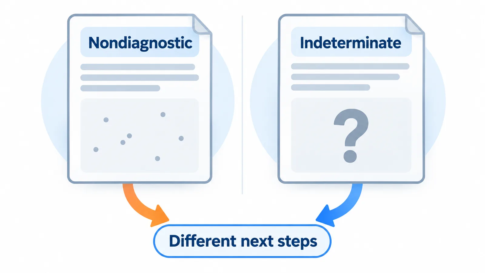
Nondiagnostic means the sample does not meet the requirements for a reliable cytologic interpretation. There may be too few suitable cells, too much obscuring material or another limitation. The label does not mean benign, malignant or “nothing was found.” It describes the adequacy of the submitted sample.
A repeat ultrasound-guided FNA is commonly considered, taking the imaging pattern and clinical context into account. Persistent inadequacy can lead to discussion of another sampling method, observation in selected circumstances or surgical assessment. The reason for the limited sample helps determine which approach would be useful.[5]
Ask whether the nodule is largely cystic, difficult to access or otherwise challenging to sample, and what would be different about another attempt. A repeat recommendation does not automatically imply a technical mistake. Some targets do not yield a useful cellular sample easily, even when the intended area is reached.
Keep the original diagnostic concern in view. If imaging is concerning, an inadequate sample has not resolved that concern. If the overall assessment is reassuring, the discussion can be different. Either way, “nondiagnostic” should lead to an explicit plan rather than be filed as a normal result.
What does Bethesda II: benign mean?
A benign result means the sampled cells have been interpreted as benign. This is reassuring and often supports observation. It should still be matched to the correct nodule and reviewed alongside the ultrasound appearance, symptoms and previous course. A reassuring word cannot resolve a mismatch by itself.
Follow-up after benign cytology depends on the imaging pattern and clinical circumstances. A high-suspicion ultrasound appearance may justify repeat assessment despite a benign sample. Conversely, repeated reliable benign findings can reduce the need for continuing cancer-focused testing. Monitoring for symptoms or important growth can remain a separate issue.[6]
Ask what the result changes for you. Does it support observation without intervention, clarify the reason for a symptom or allow a later treatment discussion? These are different outcomes. A benign label does not mean the nodule must be removed, and it does not make a persistent swallowing or voice problem irrelevant.
Bring earlier reports when a nodule has been sampled more than once. Dates, target identity and adequacy matter. Two documents with reassuring wording should not be counted as two independent confirmations without checking that each refers to the relevant lesion and an interpretable specimen.
What does Bethesda III: atypia of undetermined significance mean?
It can help to distinguish choosing a test from choosing a treatment. You may agree to further diagnostic assessment without committing to an operation. Ask whether the proposed test requires additional material, whether a previous sample can be used and how the result will be explained. These practical questions help make an uncertain category manageable without promising a particular outcome.
This category, often abbreviated AUS, means that some cell findings are atypical but do not meet the criteria for a more definitive category. It is an indeterminate result. It should not be translated into “definitely benign” or “cancer has been diagnosed.” The descriptive comment may help explain what raised uncertainty.
Possible next steps include repeat sampling, molecular testing where available, surveillance or a diagnostic surgical discussion. The choice depends on the full assessment, including ultrasound findings, the nature of the atypia, clinical history and personal priorities. Availability of a test does not make it necessary for everyone.
Molecular tests assess selected biological features in the sample and may refine risk in an indeterminate setting. A prospective follow-up study of selected Bethesda III and IV nodules with reassuring contemporary molecular results supported surveillance for many, while also showing why follow-up and study limitations matter. A reassuring molecular result is not a guarantee.[7]
Ask what the result of an additional test would actually change. If the plan would remain the same regardless, its value deserves discussion. If surveillance is chosen, establish who arranges it and what findings would prompt reconsideration. Uncertainty can be managed with a structured plan rather than an automatic operation.
What does Bethesda IV: follicular neoplasm mean?
If diagnostic surgery is proposed, ask what the team expects to learn from examining the removed tissue and how that information could affect later care. This is different from assuming that the cytology has already confirmed malignancy. You should also understand the consequences of observation or another test in your circumstances, so the decision reflects both the diagnostic concern and your priorities.
“Neoplasm” means a growth of cells; the word does not by itself establish malignancy. In this category, the cellular pattern raises a particular diagnostic question. Some distinctions depend on tissue architecture and evidence of invasion, which cannot always be established from an FNA sample alone.
The next discussion may include molecular testing and a diagnostic operation, often a lobectomy when appropriate. The decision needs the individual clinical picture. It should explain what information another test or surgery would provide, what alternatives exist and whether the proposed step would change care.
You may encounter the term NIFTP in explanations of thyroid pathology. It is a specific histologic diagnosis with strict requirements, including examination of the capsule after surgical removal. It cannot be confirmed from an FNA label alone. How such tumors are counted also affects published malignancy estimates.[8]
Do not use a reassuring ultrasound feature to erase the cytology question, or an unfamiliar pathology word to decide the extent of surgery yourself. Ask the treating specialist to connect the sample, scan and purpose of any proposed operation. The category identifies a diagnostic issue; it does not supply every detail of the answer.
What does Bethesda V: suspicious for malignancy mean?
This category means the cells raise substantial concern for malignancy, but the available features do not meet all the criteria for a definitive malignant interpretation. It needs timely specialist review. It is different from the lower-certainty atypia category and should not be treated as a routine benign result.
The descriptive part of the report matters because it may identify the type of malignancy being considered. The clinician can then decide what further assessment, review of the material or treatment planning is appropriate. Not every suspected thyroid malignancy follows the same pathway.
Ask which specialist will coordinate the next step, what additional information is required and the intended timeframe. A recommendation for surgery may be discussed, but the exact procedure cannot be chosen from the category number alone. The ultrasound map, lymph-node findings and other clinical information remain relevant.
If another opinion is sought, arrange review of the actual pathology material where appropriate, together with the full report and imaging. Repeating isolated category labels from different conversations can obscure the specific concern that needs to be addressed. The useful outcome is a coherent diagnostic and management plan.
What does Bethesda VI: malignant mean?
A malignant cytology interpretation means that the sampled cells have features diagnostic of malignancy. The report should describe the identified or suspected tumor type. This is a different situation from an indeterminate sample and requires a cancer-specific specialist pathway rather than routine benign-nodule management.
The category still does not state the cancer stage, the extent of disease or the treatment required. Those questions need appropriate clinical and imaging assessment. A small nodule, a normal hormone test or an absence of symptoms does not replace that process.
It is reasonable to ask for a clear explanation of what has been established and what remains to be assessed. Ask who is responsible for the plan and which findings could change it. Avoid applying a general benign-nodule treatment page to this result or assuming that every tumor type requires the same operation.
This article does not recommend a cancer treatment or describe selected cancer-ablation protocols. Those are separate specialist decisions. The immediate aim is to understand the report accurately and reach the appropriate team with the information needed for a considered discussion.
Why can risk estimates and next-step recommendations differ?
Keep a written timeline when reports from different laboratories are being compared. Note the date, nodule location, technique and full interpretation for each sample. A change in category can reflect a different specimen or interpretation and should be discussed rather than assumed to represent progression. If an expert review changes the wording, ask which interpretation now guides the plan and whether any additional evidence is needed.
Published percentages describe groups. They depend on the Bethesda edition, the population studied and whether only operated nodules were included. Nodules selected for surgery are not necessarily representative of every nodule with the same category. Classification changes, including how NIFTP is counted, can also alter a reported estimate.
For that reason, a percentage copied from a table should not become your personal probability. Ask which edition and local evidence the clinician is using, and whether the estimate would change the proposed next step. This guide uses the preferred 2023 category names without assigning individual risk numbers.
Imaging remains relevant after sampling. ACR’s current FAQ specifically notes that previously biopsied nodules need management informed by the biopsy result, rather than automatic application of the routine unbiopsied-nodule pathway. The report and the sample should inform each other.[9]
Useful questions are: “Does this result fit the ultrasound?”, “What uncertainty remains?”, “Would another test change the plan?” and “What are the alternatives?” A recommendation can differ legitimately when symptoms, comorbidity, local expertise or goals differ. The explanation should identify those factors instead of attributing everything to the category number.
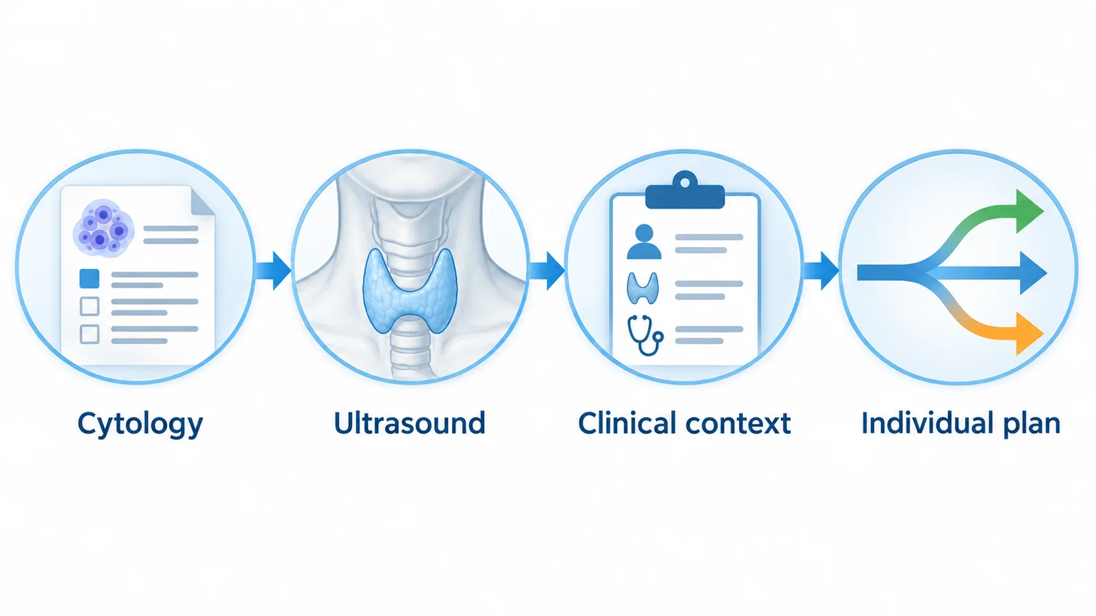
How I connect a Bethesda result with the next decision
I first confirm that the result belongs to the intended nodule and that the material was adequate for the interpretation given. I then compare the cytology with the ultrasound map and clinical concern. A reassuring word should not hide a discrepancy, and an uncertain result should not be presented as a diagnosis it has not established.
Before discussing routine benign-nodule ablation, I need reliable benign confirmation. Bethesda III, IV or V is not equivalent to that confirmation; a malignant result belongs in a separate specialist pathway. Further assessment may be the appropriate next step even when a nodule is technically accessible to a procedure.
If benignity is established, I still ask what treatment would help with. Pressure, swallowing difficulty, cosmetic concern, growth or autonomous hormone production can create different discussions. Observation and other treatment options remain relevant. My aim is to make the diagnostic evidence useful for the decision you face, without turning every biopsy result into either an automatic procedure or an unsupported promise of reassurance.[10,11]
Conclusion
Bethesda categories communicate what an FNA sample shows and how certain the interpretation is. Keep the preferred name, adequacy comment and nodule identity together. Ask how the result fits the ultrasound and what uncertainty the next step would resolve. A clear, individualized plan is more useful than a number interpreted alone.
Frequently Asked Questions
Is Bethesda the same as TI-RADS?
No. Bethesda describes the sampled cells. TI-RADS describes ultrasound appearance. The two should be interpreted together without treating their numbers as interchangeable.
Does Bethesda I mean a normal result?
No. It means the sample was nondiagnostic. Ask what limited interpretation and how the remaining diagnostic question will be addressed.
Does Bethesda II mean no follow-up of any kind?
Not automatically. It is reassuring, but the imaging pattern, symptoms and any mismatch still matter. Cancer-focused surveillance and monitoring a symptomatic nodule are different issues.
Does Bethesda III mean I have cancer?
No. It is an indeterminate result. The clinician may discuss repeat sampling, additional testing, observation or diagnostic surgery depending on the complete assessment.
Is follicular neoplasm another word for cancer?
No. Bethesda IV identifies a diagnostic question that cells alone may not resolve. Tissue architecture and other evidence can be important to the next decision.
Can molecular testing guarantee a benign diagnosis?
No. It can refine risk in selected indeterminate samples, but interpretation depends on the test, sample and clinical setting. A reassuring result may still require surveillance.
Does Bethesda V need specialist review?
Yes. It represents substantial suspicion and needs an appropriate plan. Ask who will coordinate the next step and whether additional assessment is required.
Does Bethesda VI tell me the cancer stage?
No. It identifies a malignant cytology interpretation. Tumor type, extent and management require separate specialist assessment.
Why do websites give different cancer percentages?
They may use different editions, populations or definitions. Percentages from selected groups should not be treated as an exact personal risk.
Can an indeterminate result qualify me for benign-nodule ablation?
No. It is not reliable benign confirmation. Resolve the diagnostic question before considering a routine benign-nodule treatment pathway.
Scientific References
- Ali SZ, Baloch ZW, Cochand-Priollet B, et al.. The 2023 Bethesda System for Reporting Thyroid Cytopathology. Thyroid. 2023;33(9):1039-1044. doi:10.1089/thy.2023.0141.
- Tessler FN, Middleton WD, Grant EG, et al.. ACR Thyroid Imaging, Reporting and Data System (TI-RADS): White Paper of the ACR TI-RADS Committee. J Am Coll Radiol. 2017;14(5):587-595. doi:10.1016/j.jacr.2017.01.046.
- Radiological Society of North America and American College of Radiology. Ultrasound-Guided Thyroid Biopsy. [Internet]. Accessed 2026 Oct 29.
- American Thyroid Association. Fine Needle Aspiration Biopsy of Thyroid Nodules. [Internet]. Accessed 2026 Oct 29.
- Durante C, Hegedüs L, Czarniecka A, et al.. 2023 European Thyroid Association Clinical Practice Guidelines for thyroid nodule management. Eur Thyroid J. 2023;12(5):e230067. doi:10.1530/ETJ-23-0067.
- Haugen BR, Alexander EK, Bible KC, et al.. 2015 American Thyroid Association Management Guidelines for Adult Patients with Thyroid Nodules and Differentiated Thyroid Cancer. Thyroid. 2016;26(1):1-133. doi:10.1089/thy.2015.0020.
- Kim NE, Raghunathan RS, Hughes EG, et al.. Bethesda III and IV Thyroid Nodules Managed Nonoperatively After Molecular Testing With Afirma GSC or Thyroseq v3. J Clin Endocrinol Metab. 2023;108(9):e698-e703. doi:10.1210/clinem/dgad181.
- American Thyroid Association. NIFTP: Non-invasive Follicular Thyroid Neoplasm with Papillary-like Nuclear Features. [Internet]. Accessed 2026 Oct 29.
- American College of Radiology. ACR TI-RADS FAQ [Internet]. Accessed 2026 Oct 29.
- Sinclair CF, Baek JH, Hands KE, et al.. General Principles for the Safe Performance, Training, and Adoption of Ablation Techniques for Benign Thyroid Nodules: An American Thyroid Association Statement. Thyroid. 2023;33(10):1150-1170. doi:10.1089/thy.2023.0281.
- Papini E, Monpeyssen H, Frasoldati A, Hegedüs L.. 2020 European Thyroid Association Clinical Practice Guideline for the Use of Image-Guided Ablation in Benign Thyroid Nodules. Eur Thyroid J. 2020;9(4):172-185. doi:10.1159/000508484.