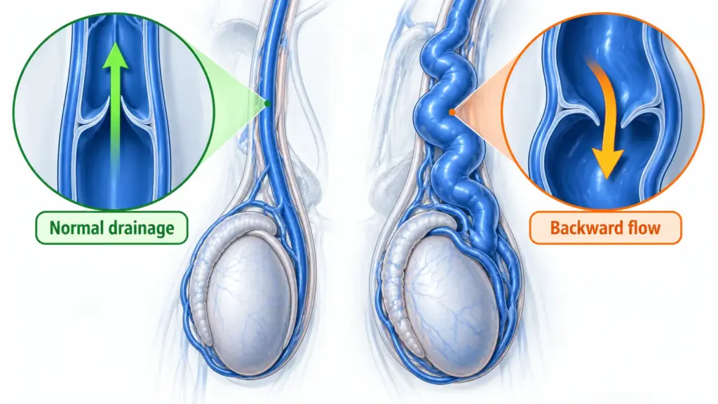
A varicocele develops when veins draining a testicle become enlarged and blood flow through that venous network is abnormal. Backward flow, called reflux, and the pressure within the drainage system help explain the finding. The veins involved are usually in the pampiniform plexus, a network around the testicle that contributes to temperature regulation.
There is rarely a single everyday action that explains why one person develops a varicocele and another does not. Anatomy matters, particularly the drainage arrangements on the left, but it is misleading to blame one workout, sexual habit, or meal. Understanding the mechanism is useful; deciding whether the finding matters clinically requires a separate assessment.
This guide separates what is known about venous drainage from proposed biological mechanisms and common causal myths. It also explains why a newly appearing or unusual finding may need further investigation without assuming that a common varicocele signals a dangerous underlying condition. In discussing causes, I separate an explanation of venous flow from evidence that the finding needs treatment.
What goes wrong with venous drainage?
Arteries bring blood to the testicle, while veins carry it away. A varicocele concerns the venous side of that circulation. Impaired drainage can allow pressure and reflux in the veins around the testicle, making them enlarged and winding. The condition is not the same as an arterial blockage and does not mean that all blood flow to the testicle has stopped.[1]
Venous valves normally help support one-way flow, but valve function is only part of the explanation. The configuration of the drainage pathway and the pressure against which blood drains also matter. It is therefore more accurate to describe a drainage problem than to tell every patient that a single broken valve is the proven cause.
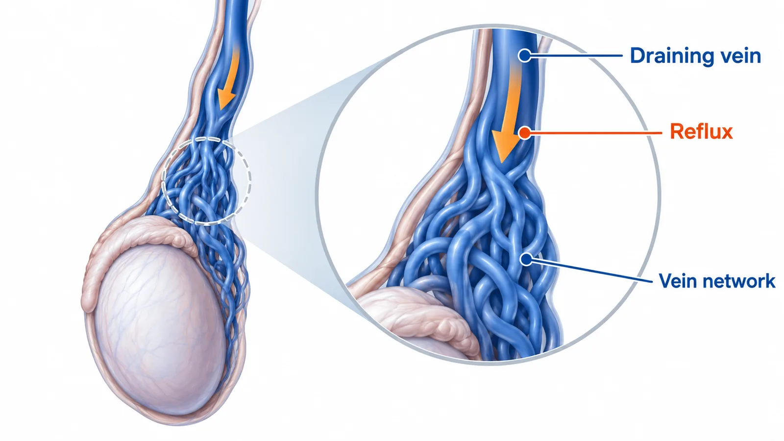
An ultrasound can demonstrate how blood moves within the veins. It cannot usually establish exactly when the condition began or identify one event responsible for it. A report describes findings at the time of the examination, rather than providing a complete history of how they developed.
Why does anatomy influence which side is affected?
The left testicular vein usually drains into the left renal vein, whereas the right usually drains directly into the main abdominal vein. Differences in the length and arrangement of these pathways help explain the predominance of left-sided findings. Individual variation also exists, including additional venous channels that may matter when planning an intervention.[2]
This anatomical explanation does not mean that the left testicle is inherently diseased or that a right-sided finding is automatically dangerous. Nor does a left-sided varicocele prove compression of the renal vein. An uncommon compression syndrome requires its own clinical assessment; it should not be inferred from a familiar anatomical diagram.
If a report mentions both sides, ask which findings were present on examination and which were visible only with imaging. The same drainage concept can apply on both sides, but their clinical significance may differ.
It is reasonable to ask whether the report actually demonstrated backward flow or only described prominent veins. Also ask whether position and straining were documented. These questions do not challenge the finding; they clarify what the examination established and what remains to be interpreted alongside your history.
How could a venous problem affect testicular function?
Research explores changes in local temperature, oxidative stress, oxygen availability, and cellular signaling. Oxidative stress means an imbalance involving reactive molecules that can affect cells. These mechanisms may help explain why some varicoceles are associated with impaired sperm production, but their relative contributions remain incompletely understood.[3]
Association does not establish that every person with a varicocele will develop a functional problem. A mechanistic explanation is also not evidence that a supplement targeting one proposed pathway will correct the venous abnormality. Patient decisions should rest on relevant clinical findings and useful outcomes, rather than the appeal of a laboratory theory.
Similarly, genetic influences under investigation do not make a varicocele a simple inherited diagnosis. A family history can be mentioned during assessment, but it does not provide a reliable individual forecast or make routine genetic testing the default for an isolated varicocele.
Do exercise, sex, or everyday habits cause it?
Discomfort becoming noticeable during activity does not prove that the activity created the varicocele. Standing and straining can change venous filling and make an existing finding easier to notice. A clinician distinguishes such triggers from an explanation for why the drainage abnormality developed in the first place.
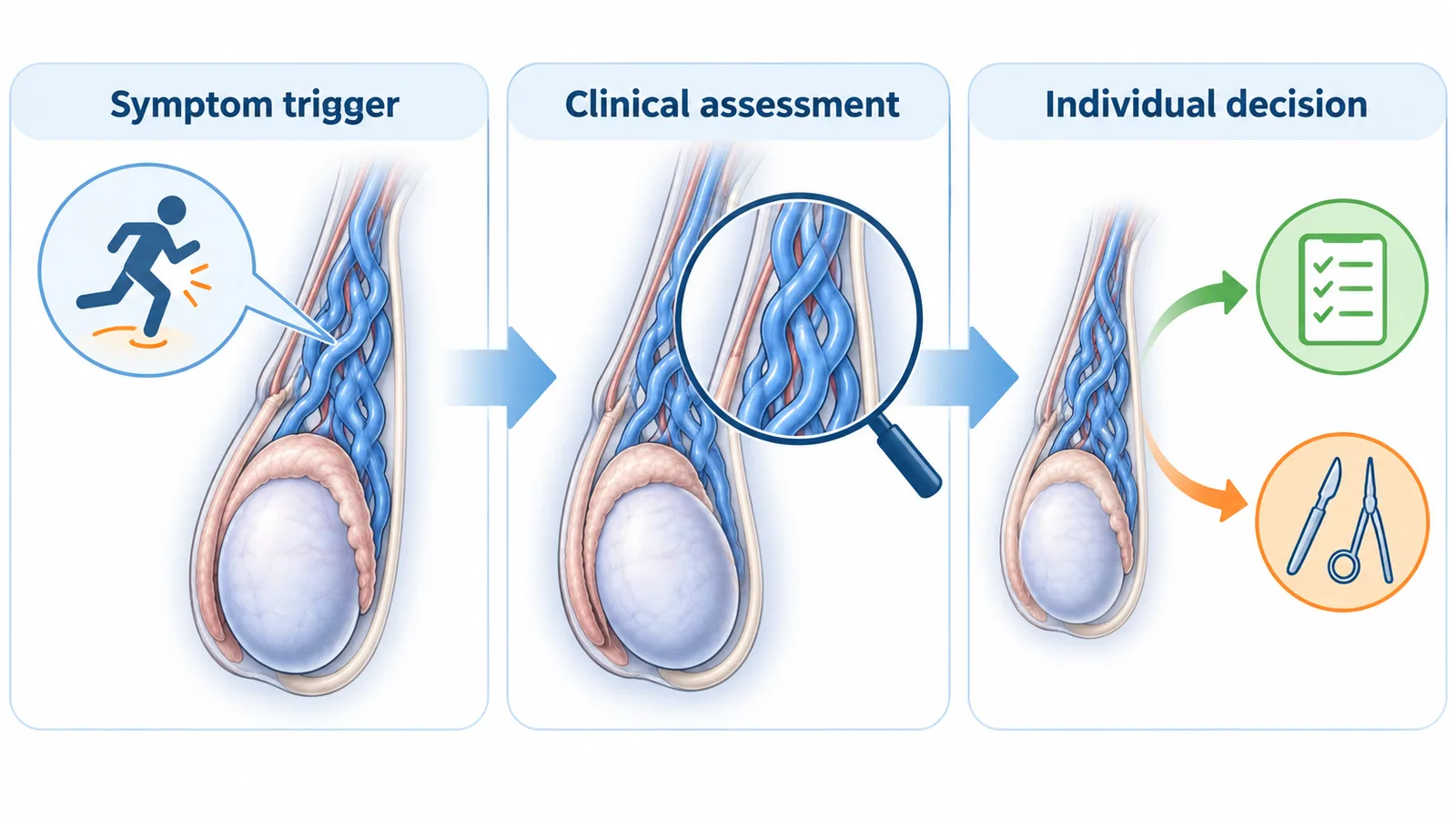
Do not turn uncertainty into a long list of prohibitions. If an activity repeatedly causes discomfort, discuss practical adjustments and assessment rather than assuming that every normal activity is harmful. Equally, do not continue through new or severe pain simply because you believe the cause is already known.
General health choices can support well-being and reproductive health, but they should not be presented as a proven method of preventing or reversing a varicocele. Ask what evidence supports any product claiming to repair valves, shrink veins, or restore fertility without an assessment.
When might another underlying problem need investigation?
Most routine varicocele discussions concern the local drainage problem. A clinician may consider further investigation when a finding is newly appearing, large, unusual, or does not reduce when lying down. Current AUA guidance does not recommend routine abdominal imaging solely because an isolated small or moderate varicocele is on the right.[4]
This is a reason to provide a clear history, not a reason to assume cancer or request a scan without clinical context. Mention when you first noticed the change, whether it varies with posture, and any other symptoms. Your clinician can decide whether a scrotal examination, Doppler study, abdominal assessment, or a different investigation addresses the concern.
Being told that a finding is longstanding can be reassuring when the rest of the assessment fits. However, an old label should not automatically explain a new lump, a different pain pattern, or a rapid change.
When reports from different years look different, ask whether the technique was comparable before assuming the condition has progressed. A change in wording, position, or measurement site may explain part of the difference. Comparing the original reports helps the clinician decide whether an apparent change is meaningful.
Does knowing the cause change what happens next?
The next step depends on whether the varicocele is clinically relevant. Observation may be appropriate when there are no meaningful symptoms or functional concerns. For selected people with infertility and a clinical varicocele, treatment can be considered, but recommendations are conditional and do not promise pregnancy. A scan finding alone is not equivalent to that clinical situation.[5]
Both surgery and embolization aim to interrupt problematic venous pathways so blood drains through other channels. Embolization uses a catheter and image guidance; it is a medical intervention with risks, including bleeding, contrast-related problems, and technical limitations. Understanding the cause does not establish that one procedure is suitable for everyone.[6]
A useful consultation question is: what evidence connects this finding with my problem? The answer may involve symptom patterns, examination, semen testing when relevant, or changes in testicular development. It should also explain what another diagnosis would mean for the plan.
My approach to understanding varicocele causes
My approach begins by separating documented findings from possible explanations. Reflux may be demonstrated, while the reason it developed or its contribution to discomfort remains uncertain. A familiar anatomical explanation can be useful without establishing a single cause for an individual patient.
I consider what the examination and any indicated Doppler study actually show: the side involved, venous flow, testicular findings, and whether another abnormality could explain the concern. These observations help keep a proposed cause from becoming a treatment recommendation on its own.
The practical discussion then turns to consequences. A symptom noticed during exercise does not establish why the veins became enlarged, and a biological theory does not prove that a supplement will correct them. Any decision about intervention needs a clinically meaningful problem and a realistic account of the expected benefit.
My clinical background provides context for a discussion about interpreting these findings and the role of image-guided treatment.
Sudden severe testicular pain needs emergency assessment rather than an appointment to discuss varicocele causes. In particular, pain with nausea or vomiting should not be attributed to a longstanding vein finding while waiting for a routine scan.[7]
I explain this distinction in how I decide whether a procedure is appropriate.
Conclusion
A varicocele is best understood as an abnormality of testicular venous drainage, influenced by anatomy and blood-flow conditions. Everyday blame and single-cause explanations are usually unhelpful. The practical question is whether the finding has clinical consequences that make monitoring or treatment worthwhile.
Frequently Asked Questions
Is a varicocele an infection?
No. It is a venous drainage problem. Infection can cause similar discomfort and needs a separate assessment when suspected.
Did lifting weights cause my varicocele?
Noticing symptoms while lifting does not establish that lifting caused it. Describe the trigger and seek assessment if discomfort persists or changes.
Does a left varicocele mean a kidney problem?
No. Left-sided anatomy helps explain the common pattern. Additional investigation depends on the history and clinical findings, not the side alone.
Can vitamins repair the veins?
They are not an established way to correct venous reflux. A product’s proposed biological mechanism does not demonstrate that it treats the underlying varicocele.
Is it definitely inherited?
No simple inheritance pattern explains every case. Research into genetic influences does not provide a reliable personal prediction or routinely require genetic testing.
Does understanding the cause mean I need treatment?
No. Treatment depends on the significance of the finding, your symptoms and goals, and the expected balance of benefits and risks.
Scientific References
- European Association of Urology. EAU Guidelines on Sexual and Reproductive Health: Male Infertility. Current online guideline; accessed 2026 Oct 29.
- Bertolotto M, Freeman S, Richenberg J, et al. Ultrasound evaluation of varicoceles: systematic literature review and rationale of the ESUR-SPIWG Guidelines and Recommendations. J Ultrasound. 2020;23:487-507. doi:10.1007/s40477-020-00509-z.
- Munoz-Lopez C, Wong A, Lewis K, Bole R, Vij SC, Lundy SD. The evolving landscape of male varicocele pathophysiology in the era of multi-omics: a narrative review of the current literature. Biology (Basel). 2024;13(2):80. doi:10.3390/biology13020080.
- American Urological Association; American Society for Reproductive Medicine. Diagnosis and treatment of infertility in men: AUA/ASRM guideline. 2020; amended 2024.
- World Health Organization. Guideline for the prevention, diagnosis and treatment of infertility. Geneva: WHO; 2025. Chapter 9: Treatment of infertility due to male factors.
- Radiological Society of North America; American College of Radiology. Varicocele embolization. RadiologyInfo.org. Reviewed 2024 Sep 1.
- National Health Service. Testicle pain. NHS; accessed 2026 Oct 29.