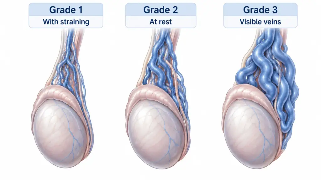
Varicocele grades 1, 2 and 3 describe how enlarged testicular veins are detected during a clinical examination. Grade 1 is felt only with straining, grade 2 is felt without straining, and grade 3 is visible as well as palpable. A finding detected only by imaging, without a palpable abnormality, is usually described as subclinical.
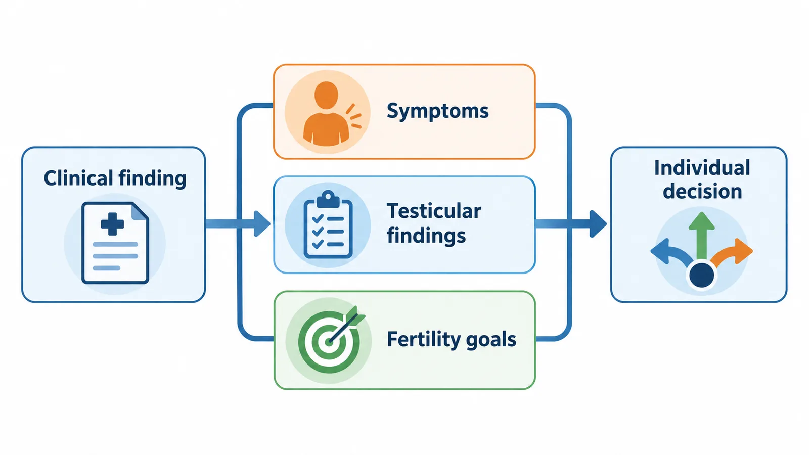
The grade is not a treatment prescription. A lower-grade clinical varicocele may matter in an infertility assessment, while a larger one may not need intervention if it causes no meaningful problem. Symptoms, testicular findings, semen results when relevant, and reproductive goals are considered together.
If your report says “grade 2” without explaining the classification, first ask whether it refers to a physical examination or an ultrasound system. Those descriptions are not automatically interchangeable. This guide explains the clinical grades and how to use them in a discussion about observation or treatment. When I interpret a grade, I ask what it adds to the clinical decision rather than treating the number as an instruction.
What do clinical grades 1, 2 and 3 describe?
The usual clinical classification is based on examining the veins around the testicle, generally while standing. For grade 1, the examiner feels the abnormal veins during a Valsalva maneuver, a brief bearing-down effort. Grade 2 can be felt at rest. In grade 3, the enlarged veins are visible and can also be felt. These are descriptions of examination findings, not three separate diseases.[1]
| Clinical description | What the examiner detects | What it does not establish |
|---|---|---|
| Grade 1 | Palpable during straining | Whether symptoms or infertility are caused by it |
| Grade 2 | Palpable without straining | An automatic need for repair |
| Grade 3 | Visible and palpable veins | A personal prediction of fertility |
The word palpable means that a trained examiner can feel the finding. It does not mean you should repeatedly check or grade yourself. The examination also evaluates the testicle and other structures, so a familiar-looking vein pattern should not explain a separate new lump.
A useful way to read the table is to focus on the examination condition, not the apparent size of an illustration. Drawings simplify anatomy and cannot show how a vein feels during examination. Two people with the same clinical grade can have different concerns and different management plans.
Is an ultrasound grade the same thing?
Not necessarily. Ultrasound classifications may combine vein location, diameter, reflux, and changes with position or straining. Some use more than three categories. A number copied from one of those systems cannot be translated directly into a clinical grade without knowing the method used.[2]
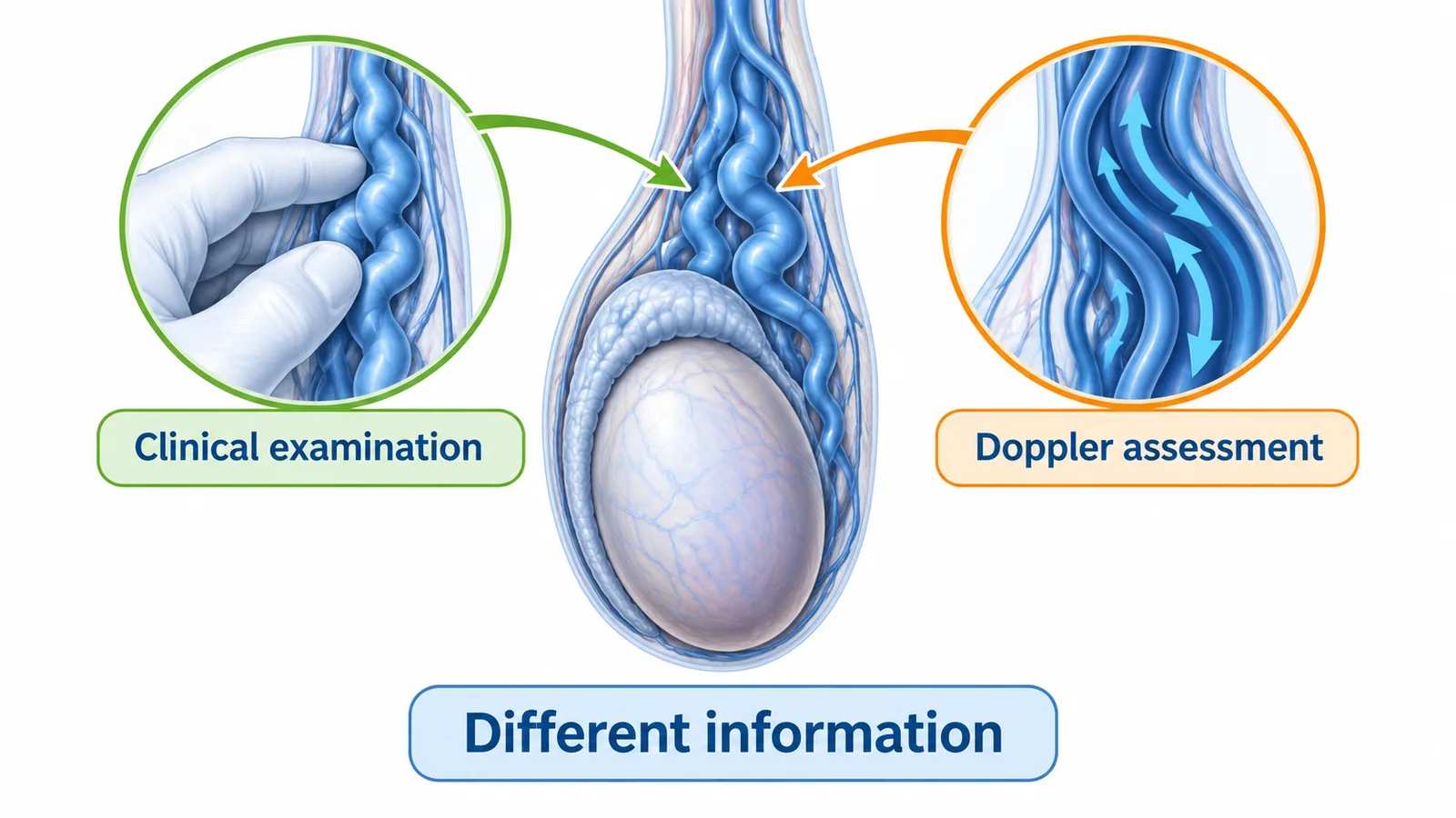
A useful report identifies the technique and records the underlying findings. For varicocele assessment, these may include the largest vein diameter under stated conditions, the presence and duration of backward flow, and testicular volume. The information can complement an examination, but does not replace its role in identifying a clinically palpable varicocele.[3]
Bring the complete reports rather than a photograph of one isolated number. The surrounding description may resolve an apparent disagreement without another investigation.
How is grade 1 different from a subclinical finding?
A clinical grade 1 varicocele can be felt during straining. A subclinical varicocele is not palpable and is detected by imaging. This distinction matters because guidelines for infertility treatment focus on clinical findings in appropriate patients. Current AUA/ASRM guidance advises against varicocelectomy for nonpalpable varicoceles detected solely by imaging.[4]
The terms small, mild, and low grade can blur this distinction if they appear without a clear explanation. Ask whether the finding was actually present on examination. Also ask whether the reason for the scan was pain, a suspected lump, infertility, or another concern; that context affects interpretation.
A decision to observe a subclinical finding is not the same as ignoring symptoms. Persistent pain or a new swelling still deserves an assessment for its cause.
Does a higher grade mean worse fertility?
A more prominent finding can be associated with impaired testicular function in studied populations, but an association does not predict an individual’s fertility. A clinical grade does not measure sperm concentration, movement, or shape. Those questions require appropriate laboratory assessment when fertility is relevant.
Semen analysis uses standardized collection and laboratory methods, and results must be interpreted with the clinical and reproductive history. A result is not a simple pass-or-fail fertility certificate. Conversely, a grade alone cannot show that sperm production is abnormal.[5]
If you are trying to conceive, explain how long you have been trying and any previous assessment for either partner. If family planning is a future consideration, ask what evaluation would be useful now and what can reasonably wait. Avoid assuming that a larger grade means an urgent procedure regardless of your circumstances.
The practical aim is to connect the finding with an outcome that matters. Improving a laboratory parameter, reducing discomfort, and achieving a pregnancy are related but distinct goals.
What else determines whether treatment is considered?
For fertility, clinicians consider whether the varicocele is clinical, whether semen parameters are abnormal, and how the couple’s situation affects the available choices. WHO guidance supports considering treatment for selected men with infertility and a clinical varicocele, but the recommendation is conditional and the certainty of evidence is low. It does not establish that every grade should be repaired.[6]
Persistent characteristic discomfort can prompt a separate discussion after other causes are considered. Testicular development and size can also matter, especially in adolescents, whose evaluation should follow an age-appropriate pathway. None of these decisions should be made from a cropped ultrasound report or a self-assigned grade.
If observation is recommended, ask what is being monitored and what change would alter the plan. If treatment is suggested, ask which specific clinical problem it is expected to improve and how uncertainty will be explained.
Does the grade choose between surgery and embolization?
No. Suitability also depends on venous anatomy, previous treatment, the intended benefit, and the balance of procedural risks and alternatives. Embolization uses a catheter to reach and close problematic venous pathways; it involves contrast and image guidance, with potential bleeding, venous injury, and other complications. Catheter access can be technically unsuccessful in some patients.[7]
Surgical repair is another option in appropriate circumstances. Comparing approaches requires a discussion of the particular technique and the outcome you hope to improve. A statement that one method “treats grade 3” is too broad to explain whether it is appropriate for you.
Ask whether the goal is symptom relief or a fertility-related change, what follow-up will assess that goal, and what happens if the varicocele persists or recurs. Neither a technically completed procedure nor a lower-looking grade guarantees the desired patient benefit.
How I decide whether a varicocele needs treatment
I begin by clarifying what the grade describes. Was the varicocele palpable during examination, and was the number assigned clinically or by an ultrasound classification? A clinical finding and an imaging-only finding can lead to different discussions, even when a report uses the same word mild.
My assessment then brings together the symptom pattern, testicular findings, and reproductive priorities. When fertility is relevant, the semen results and the couple’s circumstances add information that the grade cannot provide. If Doppler is indicated, reflux and the conditions behind the measurements matter alongside vein diameter.
The decision I want to explain is why observation, additional assessment, or an intervention would be reasonable for the particular concern. A larger visible varicocele does not automatically need repair, while a smaller clinical finding may be relevant in an appropriate infertility assessment. The intended outcome should be clear before discussing a procedure.
I also consider the limits of that reasoning. Pain may have another cause, an abnormal semen result does not establish which factor is responsible, and treatment cannot guarantee pregnancy or symptom relief. A useful plan explains what evidence supports it and what follow-up will assess.
You can review my clinical background and bring the complete examination, ultrasound, and relevant laboratory reports to that discussion.
For the wider decision framework, see how I weigh benefit, risk and alternatives.
Conclusion
Clinical grades 1, 2 and 3 describe how a varicocele is detected during examination. Ultrasound systems may describe different features. The grade helps organize a discussion, but symptoms, testicular findings, fertility assessment, and personal priorities decide whether treatment is worth considering.
Frequently Asked Questions
Is grade 1 the same as subclinical varicocele?
No. Grade 1 is palpable with straining. A subclinical finding is detected only by imaging and is not palpable.
Does grade 3 always need treatment?
No. Visibility alone does not establish a treatment indication. The clinical consequences and the likely benefits and risks matter.
Can a grade 2 varicocele affect fertility?
It can be relevant in a fertility assessment, but the grade cannot determine fertility by itself. Clinical history and appropriate semen analysis provide different information.
Why did another clinician give a different grade?
The examination conditions, observer, or classification system may differ. Compare the complete findings before concluding that the condition changed.
Does grade 1 inevitably become grade 3?
No. The grading system does not describe an inevitable sequence. Follow-up, when needed, depends on clinical circumstances rather than an assumed progression.
Can the grade choose the best procedure?
No. Anatomy, previous treatment, the intended outcome, alternatives, and procedural risks must also be considered.
Scientific References
- European Association of Urology. EAU Guidelines on Sexual and Reproductive Health: Male Infertility. Current online guideline; accessed 2026 Oct 29.
- Bertolotto M, Cantisani V, Drudi FM, Lotti F. Varicocoele. Classification and pitfalls. Andrology. 2021;9(5):1322-1330. doi:10.1111/andr.13053.
- Bertolotto M, Freeman S, Richenberg J, et al. Ultrasound evaluation of varicoceles: systematic literature review and rationale of the ESUR-SPIWG Guidelines and Recommendations. J Ultrasound. 2020;23:487-507. doi:10.1007/s40477-020-00509-z.
- American Urological Association; American Society for Reproductive Medicine. Diagnosis and treatment of infertility in men: AUA/ASRM guideline. 2020; amended 2024.
- World Health Organization. WHO laboratory manual for the examination and processing of human semen. 6th ed. Geneva: WHO; 2021.
- World Health Organization. Guideline for the prevention, diagnosis and treatment of infertility. Geneva: WHO; 2025. Chapter 9: Treatment of infertility due to male factors.
- Radiological Society of North America; American College of Radiology. Varicocele embolization. RadiologyInfo.org. Reviewed 2024 Sep 1.